The first mention of the flu was noted many centuries ago - back in 412 BC.
AD a description of a flu-like illness was made by Hippocrates. Also
flu-like outbreaks were noted in 1173. First documented
influenza pandemic that has taken a lot of
lives, happened in 1580.
In 1889-1891, a moderately severe pandemic occurred, caused by a virus of the H3N2 type.
The infamous "Spanish flu" caused by the H1N1 virus occurred in 1918-1920.
This is the most severe pandemic known.
Claimed more than 20 million lives. From "Spanish"
20-40% of the world's population has been seriously affected. Death came very
quickly. A person could still be absolutely healthy in the morning, by noon he would fall ill and
died by nightfall. Those who did not die in the first days often died from complications,
caused by the flu, such as pneumonia. An unusual feature of the "Spanish" was
that it often affected young people (usually from influenza in the first place
children and the elderly suffer).
The causative agent, the influenza virus, was discovered by Richard Shope in 1931.
Influenza A virus was first identified by British virologists Smith,
Andrews and Laidlaw (National Institute for Medical Research, London) in 1933
year. Three years later, Francis isolated influenza B.
In 1940, an important discovery was made - the influenza virus can be
cultured in chick embryos. As a result, new
opportunities for studying the influenza virus.
Taylor first isolated the influenza C virus in 1947.
In 1957-1958 there was a pandemic
Which is called "Asian flu", caused by the H2N2 virus. Pandemic
began in February 1957 in the Far East and quickly
spread throughout the world. Only in the USA during this pandemic died
more than 70,000 people.
In 1968-1969, there was a medium severity "Hong Kong flu" caused by
the H3N2 virus. The pandemic began in Hong Kong in early 1968. Most often
the virus affected older people over 65 years of age. Total number
33,800 people died from this pandemic.
In 1977-1978, a relatively mild pandemic occurred.
Called the "Russian" flu. Influenza (H1N1) virus that caused this pandemic
already caused an epidemic in the 1950s.
Therefore, those born after 1950 were the first to suffer.
Influenza pathogens belong to the family of orthomyxoviruses, which includes 3 genera of viruses influenza: A, B, C. Influenza viruses contain RNA, an outer shell that contains 2 antigens - hemagglutinin and neuraminidase, which can change their properties, especially in type A virus. Changes in hemagglutinin and neuraminidase cause the emergence of new virus subtypes that usually cause more severe and more widespread diseases.
According to the International Nomenclature, the designation of virus strains includes the following information: genus, place of isolation, isolate number, year of isolation, type of hemagglutinin (H) and neuraminidase (N). For example, A/Singapore/l/57/H2N2 refers to a virus of the genus A, isolated in 1957 in Singapore, having a variety of H2N2 antigens.
Influenza pandemics are associated with viruses of the genus A. Influenza B viruses do not cause pandemics, but local "waves" of the rise in the incidence can capture one or more countries. Influenza C viruses cause sporadic cases of the disease. Influenza viruses are resistant to low temperatures and freezing, but quickly die when heated.
Orthomyxoviruses - influenza viruses A, B, C
Structural features.
Orthomyxoviruses are enveloped (supercapsid, "clothed") viruses, the average size of virions is from 80 to 120 nm. Virions are spherical in shape. The genome is represented by a single-stranded segmented (fragmented) negative RNA. The virion has a supercapsid containing two glycoproteins protruding above the membrane in the form of protrusions (thorns) - hemagglutinin (HA) and neuraminidase (NA). Influenza A viruses have 17 antigenically distinct types of hemagglutinin and 10 types of neuraminidase.
Classification of influenza viruses is based on differences in nucleoprotein antigens (divided into viruses A, B and C) and surface proteins HA and NA. The nucleoprotein (also called the S antigen) is structurally constant and determines the type of virus (A, B, or C). Surface antigens (hemagglutinin and neuraminidase - V-antigens), on the contrary, are variable and determine different strains of the same type of virus. Changes in hemagglutinin and neuraminidase cause the emergence of new subtypes of the virus, which usually cause more severe and more widespread diseases.
The main functions of hemagglutinin:
Recognizes the cellular receptor - mucopeptide;
Responsible for the penetration of the virion into the cell, ensuring the fusion of the membranes of the virion and the cell; (Hemagglutinin provides the ability of the virus to attach itself to the cell.)
Its antigens have the greatest protective properties. Changes in antigenic properties (antigenic drift and shift) contribute to the development of epidemics caused by new Ag variants of the virus (against which herd immunity has not been sufficiently formed).
Neuraminidase responds for the dissemination of virions, together with hemagglutinin determines the epidemic properties of the virus.
Neuraminidase is responsible, firstly, for the ability of a viral particle to enter the host cell, and, secondly, for the ability of viral particles to leave the cell after reproduction.
The nucleocapsid consists of 8 segments of vRNA and capsid proteins that form a helical strand.
The life cycle of a virus.
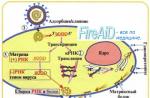
Replication of orthomyxoviruses is primarily realized in the cytoplasm of an infected cell, the synthesis of viral RNA is carried out in the nucleus. Three types of virus-specific RNA are synthesized in the nucleus on vRNA: positive messenger mRNA (a template for the synthesis of viral proteins), full-length complementary cRNA (a template for the synthesis of new negative virion RNA) and negative virion vRNA (the genome for newly synthesized virions).
Viral proteins are synthesized on polyribosomes. Further, viral proteins in the nucleus bind to vRNA, forming a nucleocapsid. The final stage of morphogenesis is controlled by the M-protein. The nucleocapsid, passing through the cell membrane, is covered first by the M-protein, then by the cellular lipid layer and supercapsid glycoproteins HA and NA. The reproduction cycle is 6-8 hours and ends with the budding of newly synthesized virions.
Antigenic variability.
(Antigenic variability of influenza viruses. The variability of the influenza virus is well known. This variability of antigenic and biological properties is a fundamental feature of influenza A and B viruses. Changes occur in the surface antigens of the virus - hemagglutinin and neuraminidase. Most likely this is an evolutionary mechanism for the adaptability of the virus to ensure survival. New strains of viruses, unlike their predecessors, do not bind specific antibodies that accumulate in the population.There are two mechanisms of antigenic variability: relatively small changes (antigenic drift) and large changes (antigenic shift).)
The modern division of orthomyxoviruses into genera (or types A, B and C) is associated with the antigenic properties of the main proteins of the nucleocapsid (nucleocapsid protein - phosphoprotein NP) and the viral envelope matrix (protein M). In addition to differences in NP and M proteins, orthomyxoviruses are distinguished by the highest antigenic variability due to the variability of the surface proteins HA and NA. There are two main types of changes - antigenic drift and antigenic shift.
Antigenic drift due to point mutations that change the structure of these proteins. The main regulator of the epidemic process in influenza is population (collective) immunity. As a result of its formation, selection of strains with an altered antigenic structure (primarily hemagglutinin), against which antibodies are less effective, occurs. Antigenic drift maintains the continuity of the epidemic process.
(Antigenic drift - occurs between pandemics in all types of viruses (A, B and C). These are minor changes in the structure of surface antigens (hemagglutinin and neuraminidase) caused by point mutations in the genes that encode them. As a rule, such changes occur every Epidemics result, as protection from previous exposure to the virus is maintained, although it is insufficient.)
However, another form of antigenic variability, antigenic shift, has also been found in influenza A viruses.(shift) associated with the change of one type of hemagglutinin (or neuraminidase) to another, i.e. on the emergence of a new antigenic variant of the virus. This is rare and is associated with the development of pandemics. In the entire known history of influenza, only a few antigenic phenotypes have been identified that cause influenza epidemics in humans: HoN1, H1N1, H2N2, H3N2, i.e. only three types of hemagglutinin (HA1-3) and two - neuraminidase (NA 1 and 2). Influenza viruses type B and C cause disease only in humans, influenza A viruses in humans, mammals and birds. The most variable influenza A viruses have the greatest epidemic role. Influenza C viruses lack neuraminidase, these viruses usually cause a milder clinical picture.
There is an opinion that antigenic shift is the result of genetic exchange (recombination) between human and animal influenza viruses. Until now, it has not been finally established where in the inter-epidemic period - outside the human population (in birds or mammals) or in the human population (due to long-term persistence, local circulation) viruses are stored that have exhausted their epidemic possibilities for a while.
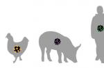
Birds are considered the primary and main hosts of influenza A viruses, which, unlike humans, have viruses with all 17 types of HA and 10 types of NA. Wild ducks are the natural hosts of influenza A viruses, in which the pathogen is located in the gastrointestinal tract and does not cause significant damage to the owners. Viruses show their pathogenic properties when they pass to other birds and mammals. Among mammals, the greatest importance is attached to pigs, which are considered an intermediate host and compared with a “mixing vessel”.
(Modern human influenza viruses are weakly transferred to animals. All influenza A pandemics since 1930 began in China, the main gateway of distribution is Siberia (mass bird migrations).
H1N1 - 1930 Identified in humans, pigs, whales (1972), domestic and wild birds. It is associated with the famous Spanish flu pandemic. This type has been re-introduced since 1977.
H2N2 has been detected since 1957. in humans and birds. Epidemics associated with these viruses came periodically. Now both types are identified in parallel.
H3N2 was identified in 1963. (Hong Kong).
Virus A/ Singapore/1/57 (H2N2) has three genes from Eurasian avian influenza viruses, virus A/ Hong Kong/1/68 (H3N2) contains 6 genes from Singapore virus and two from birds. These data confirm that humanity receives new epidemic types of influenza A viruses from birds - the primary host. The nearest forecast is the possibility of the emergence of new epidemic variants of the influenza A virus that have hemagglutinin HA5 or 7 (it is enough to replace one or two amino acids in their structure).)
A feature of influenza viruses is their ability to variability (the variability of the influenza virus). Small changes in the antigenic structure of viral proteins are called antigenic drift (from the English word drift - slow flow). Such changes occur in influenza viruses every year, and for this reason, influenza epidemics occur every year.
Influenza virus changes.
Sometimes there are more significant changes in the antigenic structure of the influenza virus, which are called shift (from the English word shift - jump). As a result of this variability, influenza viruses with completely new properties appear, and therefore the entire population of the planet is susceptible to infection. Such viruses cause influenza pandemics. Dramatic changes in the genome of the influenza virus occur every 20-40 years and, as a rule, as a result of gene reassortment, that is, the exchange of genes between human and animal influenza viruses. It is believed that this is how the new influenza virus A / California / 4/2009 (H1N1) appeared, which caused an influenza pandemic on the planet in 2009. Special studies have shown that the causative agent of this influenza pandemic is a complex reassortant, which included genes from the classic swine influenza virus, genes from the swine influenza virus of the Eurasian line, passed to them from the bird virus in the late 70s, and genes from the virus swine influenza of the North American line, which in turn is a reassortant and includes a gene from the human influenza A (H3N2) virus.
Since the summer of 2011, a new variant of the influenza A(N3N2) virus has been isolated from some influenza patients in the United States, which differs in its genetic characteristics from the currently circulating seasonal influenza A(H3N2) virus. This variant of the influenza virus is referred to as influenza A(H3N2)v.
Experts carefully monitor the circulation of influenza viruses in various regions of the world. This is necessary for recommendations on the composition of influenza vaccines and forecasting the epidemiological situation.
Is the swine flu pandemic over?
According to the World Health Organization, published in August 2010, the new virus A(H1N1)pdm09 has largely completed its cycle of development. The world has entered a post-pandemic period. But that doesn't mean the new A(H1N1)pdm09 virus is gone. Experts believe that it will behave like a seasonal flu virus. This virus will still cause disease. Therefore, precautions must be taken to reduce the risk of infection.
You can also read on this topic:A virus is a precellular form of living matter.
Characterized by:
The absence of a cell wall
The presence of one nucleic acid (RNA or DNA)
Often, complication in the form of opportunistic processes
Blood reaction in the form of leukopenia, secondary immunodeficiency
Insensitivity to antibiotic therapy.
FLU(Family - Ortomyxoviridae)
Acute viral respiratory infection.
Periodically spreads in the form of epidemics and pandemics. History reference:
the first pandemic "Spanish flu", in 1818-1820 of the last century, type A subtype H1N1.
The second is 1957-1960, "Asian" highlighted in Singapore. type A subtype H2N2
Third 1968-1970 "Hong Kong" type A subtype H3N2
It is characterized by phenomena of general intoxication, fever, damage to the upper respiratory tract, nervous and cardiovascular systems.
The structure of the influenza virus type A virion
It has a spherical shape, the size is 80-120 nanometers.
A complex RNA-containing virus consisting of a central part of the genome (core) and a supercapsid.
The virus genome is a helical, single-stranded, fragmented negative RNA, consisting of 8 segments that encode 10 viral proteins. RNA fragments have a common protein shell that combines them to form a ribonucleoprotein (RNP).
Surrounding the RNP is a layer of matrix protein (M-layer), which gives the virion strength.
On top of the M-layer, adjacent to it, is located supercapsid- lipoprotein membrane of cellular origin with embedded virus-specific glycoproteins hemagglutinin (H), named for their ability to agglutinate erythrocytes, and neuraminidase(N), an enzyme protruding from the surface of the virion in the form of many spines.
Hemagglutinin provides the ability of the virus to attach to the cell. Neuraminidase is responsible, firstly, for the ability of a viral particle to enter the host cell, and, secondly, for the ability of viral particles to leave the cell after reproduction.
The nucleoprotein (also called the S antigen) is structurally constant and determines the type of virus (A, B, or C). Surface antigens (hemagglutinin and neuraminidase V antigens), on the contrary, are variable and determine different strains of the same type of virus.
Influenza virus antigens
Internal antigens(S-antigen) - these are antigens of nucleocapsid and matrix protein proteins (NP-protein and M-protein) type-specific antigens They are stable, type-specific, and do not give cross-reactions.
NP-protein binds Co and is determined by RSK
Surface antigens(V-antigens) - are protective antigens - these are antigens of surface glycoproteins - hemaaglutinin (H-antigen) and neuraminidase (N-antigen). ANTs to these ANGs have a virus-neutralizing property. Investigate in RTGA (ANGv + Er + ANTv).
Superficial ANGs are distinguished by their diversity and variability (15 variants of H-antigens, 10 N-antigens). The human influenza virus is characterized by hemagglutinins H1, H2, H3 and neuraminidase N1 and N2. Various variants of the combination of H- and N-antigens determine the subtypes of the type A virus, for example. A/H1N1/, A/H2N2/ and A/H3N2.
Antigenic variability driven by 2 processes:
Drift antigens- minor changes in the structure of H-N antigens due to point mutations in genes , who encode them. As a result, altered serovariants (strains) of the same subtype are formed.
Shift antigens-complete replacement of the genome region encoding the synthesis of H- and N-antigens, leading to the formation of a new influenza virus subtype.
Epidemiology
Flu:
Anthroponous ARVI
Highly contageous
Transmission mechanism - aerogenic
Transmission route - airborne
Intensive reproduction of viruses occurs in the upper respiratory tract, so a short incubation period - from several hours to 2 days
The ability to antigenic variability - drift and shift.
The epidemic spread of the disease is ubiquitous.
The peak incidence increases in the autumn-winter period
Influenza virus reproduction
Adsorption of viruses on susceptible cells
Entry of virions into the cell
Transportation of the virus to the cell nucleus and further deproteinization
Expression of the viral genome and synthesis of virion components
Formation of virions and their exit from the cell
The reproduction cycle of the influenza virus lasts 6-8 hours. The infected cell does not die immediately and can produce several thousand virions.
Pathogenesis
The influenza virus selectively infects the epithelium of the respiratory tract (mainly the trachea). Reproducing in the cells of the cylindrical epithelium, it causes their degenerative changes, using the contents of the epithelial cells to build new viral particles. A massive release of mature viral particles is often accompanied by the death of epithelial cells, and necrosis of the epithelium and the associated destruction of the natural protective barrier leads to viremia. Influenza virus toxins, together with the decay products of epithelial cells, have a toxic effect on the cardiovascular, nervous (central and autonomic) and other body systems. Influenza infection leads to immune suppression, and when the secondary bacterial flora is introduced through the necrotic surface of the respiratory mucosa, various complications can occur.
In the pathogenesis of influenza, five main phases of the pathological process are distinguished:
reproduction of the virus in the cells of the respiratory tract;
viremia, toxic and toxic-allergic reactions;
damage to the respiratory tract with the predominant localization of the process in any part of the respiratory tract;
possible bacterial complications from the respiratory tract and other body systems;
reverse development of the pathological process.
In the basis of the defeat of various organs and systems in influenza, the leading role is played by circulatory disorders, the cause of which are violations of the tone, elasticity and permeability of the vascular wall, primarily capillaries. An increase in the permeability of the vascular wall leads to a violation of microcirculation and the occurrence of a hemorrhagic syndrome (nosebleeds, hemoptysis, and in severe cases, hemorrhages into the substance and membranes of the brain, into the alveoli, which is manifested by the syndrome of infectious-toxic encephalopathy or hemorrhagic toxic pulmonary edema).
flu causes decreased immunological reactivity. This leads to an exacerbation of various chronic diseases, as well as to the occurrence of secondary bacterial complications. The most common and serious complication of influenza is acute pneumonia. It is now generally accepted that influenza pneumonia is of a mixed viral-bacterial nature, regardless of the timing of its occurrence.
Clinical forms of influenza
uncomplicated form
Light degree
Moderate severity
Severe degree
Complicated form
Light degree
Moderate severity
Severe degree
Extremely severe (hypertoxic) degree
Immunity
Permanent factors of nonspecific protection (cellular and humoral): excretory function of the body, serum inhibitors, alpha-interferon, secretory IgA.
Factors induced by the virus (non-specific - an increase in body T, and specific)
Persistent post-infectious cellular and humoral immunity, which is distinguished by its narrow type, subtype, variant specificity and is directed against the serovariant (strain) of the influenza virus that caused a certain disease.
Therefore, anti-influenza immunity is subtype- and strain-specific.
Microbiological diagnostics is based on:
Isolation and identification of the virus
Determination of viral ANG in the patient's cells
Search for virus-specific ANTs in the patient's serum.
Material for research: nasopharyngeal discharge, smears - prints from the nasal mucosa, post-mortem examination of autopsies
Laboratory diagnosis of influenza- early and retrospective - carried out to confirm the clinical diagnosis, differentiate influenza from SARS of another etiology and for epidemiological purposes.
Early diagnosis : in the first 3 days and no later than the 5th day of illness, ANG of influenza viruses is detected using express methods Material under study: mucus from the nasal passages and nasopharynx, taken with tampons, by washing, by the method of smears-imprints from the mucous membrane of the lower nasal conchas, as well as sectional material after their special processing.
Express Diagnostics: 2-5 hours, more often RIF (direct and indirect). Specific ANG of the influenza virus and intracellular inclusions are detected by their bright emerald green glow in the cytoplasm and nucleus of infected epithelial cells.
Virological method
Virus isolation in chick embryos:
A combined infection of 10-11 day old embryos with the test material is carried out in the amniotic and allantoic cavities. After 3 days of incubation att 35C, the presence of viruses in the amniotic and allantoic fluid is checked using RHA with erythrocytes of chickens, guinea pigs or humans, and the virus titer is established.
Next, serological identification of the isolated virus is carried out using RSK to determine the type of virus (A, B or C) and RTGA to establish the subtype or strain of the influenza virus. Reactions put with the appropriate diagnostic sera.
Isolation of viruses in cell cultures
It is carried out by infecting several types of cell cultures, primary cultures of human kidneys and some animals are more often used. Cell cultures are incubated for a week at T-33C, daily recording changes in the cell monolayer in order to detect CPP (RTGA, RGA, IF method). Next, chicken embryos are infected with a virus-containing cultural liquid, an allantoic liquid with a high content of viruses is obtained, and the isolated virus is identified.
Retrospective diagnosis of influenza (serological study)
Serological examination of paired sera taken at the onset of the disease and after 7-14 days. An increase in the titer of specific ANTs during the course of the disease by at least 4 times (in young children - 2 times) makes it possible to establish the exact etiology of influenza.
Specific prophylaxis
Vaccination:
Whole virus vaccines (1st generation) - inactivated and live.
Split-split vaccines (2nd generation) contain internal and external ANG of influenza viruses and do not contain lipids removed after treatment of virions with solvents or detergents.
Subunit vaccines (3rd generation) are the most purified, contain external H- and N-antigens of influenza viruses
Emergency Prevention:
It is carried out during the epidemic rise in the incidence. Distinguish planned prevention organized in children's institutions, work collectives and focal in families of influenza patients. For emergency prophylaxis, antiviral drugs are used, sometimes immunoglobulin prophylaxis is carried out.
Flu treatment
Antiviral drugs are used to treat influenza:
Interferon preparation(influenza A virus + antitoxic effect in influenza B)
Remantadine(influenza virus A + B) inhibits the synthesis of the M-protein, which leads to disruption of the reproduction cycle and prevents the formation of full-fledged virions.
Arbidol and amixin, are interferon inducers and immunomodulators that affect all types of influenza viruses
In severe forms of influenza in the first 3 days of illness, the introduction is indicated anti-influenza immunoglobulin.
symptomatic treatment. In the presence of bacterial complications, antibiotics and sulfonamides are prescribed.
Anergy clonal- the state of functional unreactivity of individual clones of lymphocytes for specific antigens.
Antigen-antibody complex (immune complex) is the product of an antigen-antibody reaction. It is of great importance in the pathogenesis of many diseases.
Antigen-antibody reaction- specific interaction of the antigen with the corresponding antibody.
Antigenic variability- a change in specific surface antigens of an organism within a biological species. Most intensely manifested in influenza viruses.
Antigenic modulation- the disappearance of surface antigens under the influence of antibodies.
Antigenic specificity- structural features that distinguish a certain antigen from the individual, antigenic composition of the immunized organism; antigenic specificity does not imply the ability of carriers of this specificity to induce an immune response; haptens have antigenic specificity, reacting with pre-existing antibodies, but they themselves cannot cause their formation. Antigenic specificity gives the antigen the ability to selectively react with specific antibodies or sensitized lymphocytes.
antigenicity- the ability of a substance to interact specifically with the products of the immune response.
Antigenic determinants (epitopes)- specific parts of the antigen molecule, to which specific antibodies are produced, and with which the products of the immune response react.
Antigenic drift- a gradual change in the antigenic specificity of virus structures (HA and NA), occurring over several years, due to spontaneous point mutations.
Antigenic shift- changes in the entire antigenic structure of hemagglutinin or neuraminidase. This process leads to the emergence of new virus subtypes. Antigenic shift is based on the mechanisms of genetic recombination between individual virus subtypes.
Antigen presenting cells- highly specialized cells capable of absorbing and processing antigens, as well as presenting peptide antigenic fragments on the cell surface in combination with MHC class I or II molecules; main antigen-presenting cells: macrophages, dendritic cells, B-lymphocytes.
Antigen-recognizing B-cell receptor (surface immunoglobulin - sIg)- surface monomeric form of immunoglobulin belonging to the IgM class; able to interact with a free antigen not associated with any additional molecules.
Antigen recognition T-cell receptor (TCR)-heterodimer expressed on the surface of T-cells in complex with single-domain C3 proteins; the main function is the recognition of an immunogen (antigenic peptide + MHC class I or II molecules) on the surface of an antigen-presenting or virus-infected cell.
Antigens- Substances that induce an immune response.
Histocompatibility antigens (HLA) are antigens encoded by the major histocompatibility complex. Histocompatibility antigens contribute to the recognition of foreign antigens, play a decisive role in the cooperation of immunocompetent cells during the development of humoral and cellular immunity, and are the main structures in the implementation of transplantation immunity reactions. Represented by molecules of two classes.
Synthetic antigens artificially synthesized antigens.
T-dependent antigens- antigens, the development of an immune response to which requires the participation of helper T-lymphocytes.
Antigens T-independent- antigens, the development of an immune response to which does not require the participation of helper T-lymphocytes.
Antiserum- serum containing specific antibodies.
Antibodies- immune proteins that are formed in the body in response to the intake of an antigen and have the ability to specifically interact with it.
Antibodies immune- antibodies produced by the body as a result of an infection or developed in response to immunization with an antigen.
Monoclonal antibodies- antibodies produced by a single clone of antibody producers. These are antibodies of one class, subclass and one specificity. Antibodies usually produced in the body in response to antigenic stimulation are the products of activity of several clones of antibody producers.
Antibodies are normal- antibodies contained in the serum of healthy individuals, the production of which is not associated with past infection or any immunization.
Anticoagulants - drugs for the prevention of blood clots in the island with atrial fibrillation.
Antibody-dependent cellular cytotoxicity (ADCC)– destruction of target cells coated with antibodies by effector cells with an Fc receptor.
Influenza A/H1N1 as a typical emerging infection: General characteristics of influenza viruses, variability, emergence of new pandemic strains
Influenza viruses - RNA-containing viruses - belong to the family. Orthomyxoviridae and are divided into viruses A, B, and C (Table 1).
Table 1.
Comparative characteristics of influenza viruses
| Criteria | Type A | Type B | Type C |
| Disease severity | ++++ | ++ | + |
| natural reservoir | There is | Not | Not |
| human pandemics | calls | Does not cause | Does not cause |
| Human epidemics | calls | calls | Does not cause (only sporadic disease) |
| Antigenic changes | Shift, drift | Drifting | Drifting |
| Segmented genome | Yes | Yes | Yes |
| Sensitivity to rimantadine | sensitive | Not sensitive | Not sensitive |
| Susceptibility to zanamivir | sensitive | sensitive | - |
| Surface glycoproteins | 2 (HA, NA) | 2 (HA, NA) | 1(HA) |
The influenza virus has a spherical shape and a size of 80-120 nm. The core is represented by a single-stranded negative RNA chain, consisting of 8 fragments that encode 11 viral proteins.
Influenza A viruses are widely distributed in nature and affect both humans and a wide range of mammals and birds. Influenza viruses types B and C have been isolated from humans only.
Epidemiologically significant are 2 subtypes of influenza A virus - H3N2 and H1N1 and influenza virus type B (A.A. Sominova et al., 1997; O.M. Litvinova et al., 2001). The result of such co-circulation was the development of influenza epidemics of various etiology in the same epidemiological season in different countries. The heterogeneity of the population of epidemic viruses also increases due to the divergent nature of the variability of influenza viruses, which leads to the simultaneous circulation of viruses belonging to different evolutionary branches (O.M. Litvinova et al., 2001). Under these conditions, prerequisites are created for simultaneous human infection with various pathogens, which leads to the formation of mixed populations and reassortment both between viruses of co-circulating subtypes and among strains within the same subtype (O.I. Kiselev et al., 2000).
Classification of influenza virus types is based on antigenic differences between two surface glycoproteins, hemagglutinin (HA) and neuraminidase (NA). According to this classification, influenza viruses are divided into 3 types - influenza viruses type A, type B and type C. There are 16 subtypes of HA and 9 subtypes of NA.
Rice. 1. Classification of influenza A viruses and species of animals and birds - intermediate and final hosts in the chain of transmission of infection to humans.
Recently discovered subtype 16 (H16) of hemagglutinin
Note: ∗ HA 7 and NA 7-NA8 were also found in horses
On fig. 1 shows subtypes of influenza A viruses and their intermediate hosts and natural reservoirs (migratory birds). The main hosts of influenza A viruses are those species that are susceptible to influenza.
In the human population, influenza A viruses of only three subtypes with HA1, HA2 and HA3 have been identified so far. At the same time, viruses contain only two types of neuraminidase - NA1 and NA2 (Fig. 1). Their stable circulation has been proven over the past century since the 1918 pandemic (R.G. Webster et al., 1978; K.G. Nicholson et al., 2003).
Influenza A viruses (to a lesser extent B) have the ability to change the structure of HA and NA. Influenza A virus is characterized by two types of variability:
- point mutations in the viral genome with a corresponding change in HA and NA (antigenic drift);
- complete replacement of one or both surface glycoproteins (HA and NA) of the virus by reassortment / recombination (antigenic shift), which results in a fundamentally new version of the virus that can cause influenza pandemics.
For the influenza B virus, antigenic variability is limited only by drift, tk. it does not seem to have a natural reservoir among birds and animals. The influenza C virus is characterized by a high stability of the antigenic structure and only local outbreaks and sporadic cases of the disease are associated with it.
Of some interest emergence of new strains of the influenza virus in the human population and associated pandemics (Fig. 2). On fig. Table 2 shows the main antigenic shifts associated with pandemics of the 20th century caused by influenza A viruses:
- in 1918 the pandemic was caused by the H1N1 virus;
- in 1957 - H2N2 strain A/Singapore/1/57;
- in 1968 - H3N2 strain A/Hong Kong/1/68;
- in 1977 - H1N1 strain A / USSR / 1/77 (many scientists did not consider this a pandemic, but with the advent of this strain, a situation arose with the simultaneous co-circulation of 2 strains of the influenza A virus - H3N2 and H1N1).
In 1986, the A/Taiwan/1/86 virus caused a massive influenza A/H1N1 epidemic in China that lasted until 1989. Drift variants of this virus survived until 1995, causing localized outbreaks and sporadic cases. According to the results of molecular biological studies, multiple mutations arose in the A/H1N1 virus genome in these years. In 1996, two antigenic variants of the A/H1N1 influenza virus appeared: A/Bern and A/Beijing, their feature was not only antigenic, but also geographical disunity. Thus, in Russia, the A/Bern influenza virus took an active part in the influenza epidemic of 1997-98. In the same season, circulation of A/Beijing virus strains was registered in the east of the country. Later in 2000-2001. Influenza A/H1N1 virus has become the causative agent of the influenza epidemic in Russia. Modern A/H1N1 influenza viruses have low immunogenic activity; freshly isolated virus isolates interact only in mammalian erythrocytes (group 0 human and guinea pigs).

Rice. 2. Emergence of new strains of influenza virus in the human population and associated pandemics
During the past century, influenza A viruses have undergone significant genetic changes, resulting in global pandemics with high human mortality. The biggest influenza pandemic (H1N1) was in 1918-1919. ("Spaniard"). The virus, which appeared in 1918, made a pronounced drift, its initial (Hsw1N1) and final (H1N1) variants are considered shift. The virus caused a devastating epidemic that claimed 20 million lives (half of the dead were young people aged 20 to 50 years (M.T. Osterholm, 2005).
Research J.K. Tanbenberger et al., (2005) showed that the virus that caused the 1918 pandemic was not a reassortant between the avian influenza virus and the human influenza virus - all 8 genes of the H1N1 virus were more similar to variants of the "avian" virus than to the human one (Fig. .3). Therefore, according to R.B. Belshe (2005), an avian influenza virus, should infect (bypassing the intermediate host) humans, being transmitted from person to person.

It is important to note that avian influenza viruses "participate" in the emergence of new "human" influenza viruses, which are characterized by high pathogenicity and the ability to cause pandemics (EG Deeva, 2008). These viruses (H1N1, H2N2 and H3N2) had a different set of internal genes, the origin of which indicates their phylogenetic relationship with avian and swine viruses.
What are the mechanisms of origin of pandemic strains and what biological characteristics are necessary for the emergence of a highly pathogenic virus with pandemic potential?
Influenza A viruses are characterized by a high frequency of occurrence of reassortants as a result of mixed infection, which is due to the segmentation of the viral genome. The predominance of a reassortant of a certain gene composition is considered the result of selection, in which the one that is most adapted to reproduction under given conditions is selected from an extensive set of different reassortants (N.L. Varich et al., 2009). The strain-specific properties of genomic segments can have a strong influence on the gene composition of reassortants under non-selective conditions. In other words, the hallmark of influenza viruses is that eight of the gene segments, especially the HA gene, mutate frequently and unpredictably. Reassortment plays an important role in the emergence of new virus variants, in particular in the origin of pandemic strains. And sometimes the possibility of the emergence of a virus with higher virulence during a pandemic cannot be ruled out.
Modern studies have shown that the gene structure of the new A/H1N1 virus is complex and, as we noted in the introduction, it includes the genes for swine flu that infects pigs in North America; swine flu genes affecting pigs in Europe and Asia; bird flu genes; human influenza genes. Essentially, the genes for the new virus come from four different sources. A micrograph of influenza A/H1N1 virus is shown in fig. 4.

Rice. 4. Micrograph of influenza A/H1N1 virus
WHO has published "Guidelines for Influenza Laboratories" and presented new data on the sequence of viral genes and their length of the reassortant new influenza A / H1N1 virus (isolate - A / California / 04/2009): HA, NA, M, PB1, PB2, PA, NP, NS. These data indicate the formation of a new pandemic variant of the virus, creating a general vulnerability to infection due to the lack of immunity. It becomes clear that pandemic variants of the influenza virus arise through at least two mechanisms:
- reassortment between animal/avian and human influenza viruses;
- direct adaptation of the animal / bird virus to humans.
To understand the origin of pandemic influenza viruses, it is important to study the properties of the natural reservoir of infection and the evolutionary paths of this family of viruses during host change. It is already well known and arguable that waterfowl are the natural reservoir of influenza A viruses (adapted to these intermediate hosts over many centuries), as evidenced by the carriage of all 16 HA subtypes of this virus. Through the faeces of birds, which can remain in water for more than 400 days (Avian influenza ..., 2005), viruses can be transmitted to other animal species when drinking water from a reservoir. (K. G. Nicholson et al., 2003). This is supported by phylogenetic analysis of the nucleic acid sequences of different subtypes of influenza A viruses from different hosts and from different geographic regions.
Analysis of the nucleoprotein gene sequences showed that avian influenza viruses evolved with the advent of 5 specific host lines: viruses of wild and domestic horses, gulls, pigs and humans. Moreover (!) human and swine influenza viruses constitute the so-called sister group, which indicates their close relationship and, of course, a common origin. The precursor to human influenza viruses and the classic swine virus appear to have been entirely 'avian' in origin. In the countries of Central Asia, for obvious reasons, pork is not popular, and these animals are practically absent in animal husbandry. This leads to the fact that (unlike China, for example), this region does not have the main intermediate host in the population of domestic animals - pigs, so the probability of "birth" of pandemic viruses in the Central Asian region is lower than in China, which practically follows from data on the analysis of their origin (Avian Influenza, 2005). A permanent source of pandemic influenza virus genes exists (in a phenotypically unchanged state) in the natural reservoir of waterfowl and migratory bird viruses (R.G. Welster, 1998). It should be borne in mind that the precursors of the viruses that caused the Spanish flu pandemic (1918), as well as the viruses that were the source of the genome of the Asia / 57 and Hong Kong / 68 pandemic strains, are still circulating among the population of wild birds with minor mutational changes (Influenza birds…, 2005).
Comments
(visible only to specialists verified by the editors of MEDI RU)